

Visualizing Visceral Fat
Visceral fat can easily be visualized on routine imaging studies, particularly on CT (computed tomography) and MRI (magnetic resonance imaging), both of which can easily differentiate visceral fat from subcutaneous fat.
Given the enormous clinical implications of visceral obesity, one would think that identifying visceral fat on imaging studies would be a priority. Instead, the presence of visceral fat is almost never mentioned in radiology reports.
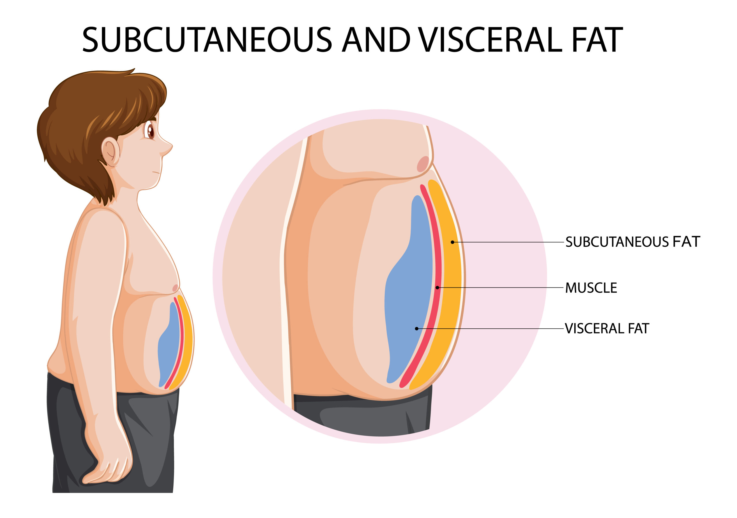
Identifying visceral fat
Recall that visceral fat is beneath the abdominal muscle layer and contributes to the protrusion of the abdomen. In contrast, subcutaneous fat is located outside the abdominal cavity and is the “pinchable” fat.
Here’s a useful visualization of visceral fat enveloping the abdominal organs.
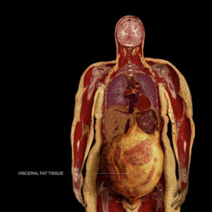
Visceral fat on CT
Note: Permission granted to use these images.
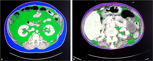
The 3 sets of paired images below are from patients I have cared for.
Patient A (left image) is a man in his 30’s with a BMI of approximately 24. He has no evident metabolic disease.
Patient B (right image) is a man in his 70’s with a BMI of 42. Aside from obesity, he suffers from type 2 diabetes complicated by vascular disease, including heart disease and peripheral vascular disease, kidney disease, and neuropathy (nerve damage). He also has other manifestations of poor metabolic health, including obstructive sleep apnea, hypertension (high blood pressure), gastroesophageal reflux disease (GERD), an enlarged prostate, high triglycerides, low HDL cholesterol, and heart failure. He is on an extremely high dose of insulin, further contributing to his insulin resistance.
Transverse Plane
The transverse plane is the standard method by which CT images are interpreted. For reference, imagine yourself standing at the foot of the bed and looking upward at slices of the patient’s body in cross-section.
![]()
For reference, visceral fat appears dark gray on CT images, often appearing to have visible streaks coursing through it – prominently displayed in the image on the right. Bone appears bright white, and muscles appear light gray. Black represents air.
Note Patient A’s abdomen has very little visceral fat, with most of the space taken up by muscles and loops of intestine. In contrast, Patient B has a large amount of visceral fat seen infiltrating the entire abdomen amidst the loops of intestine, along with relatively small muscle mass.
To better visualize visceral fat on CT, in the following example, visceral fat has been colored green, while subcutaneous fat has been colored blue.
Coronal Plane
The coronal plane is a reconstruction of the original CT images (slices) with a front-to-back perspective.
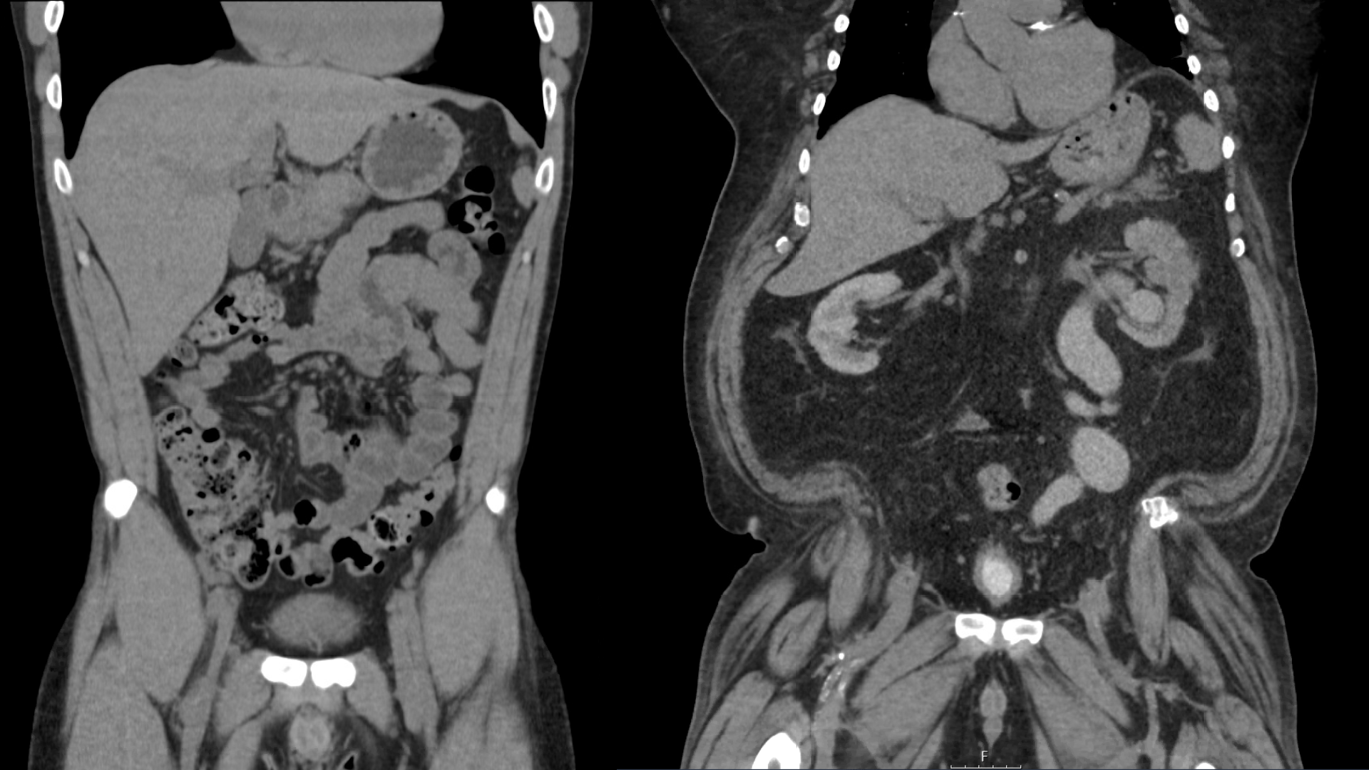
From this perspective, one can appreciate the difference in muscle tone, muscle mass, and overall burden of visceral fat. Patient B on the right appears to have increased pressure within the abdomen causing protrusion of the abdominal cavity in all dimensions. Note also the calcification in Patient B’s heart indicating the presence of coronary artery disease (heart disease).
Sagittal Plane
The sagittal view is a reconstruction of the original CT images (slices) that allows a whole-body view from the side perspective.
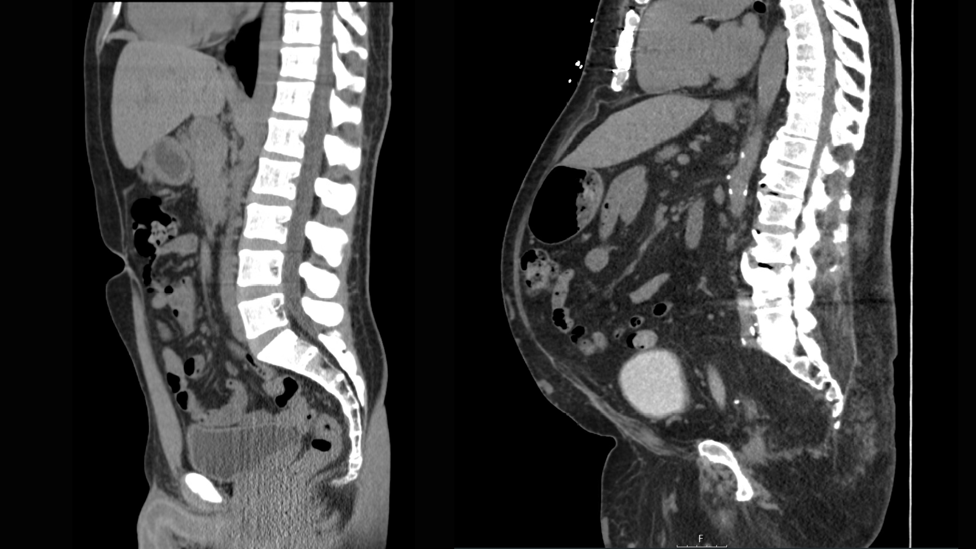
While Patient A and Patient B appear to have relatively similar amounts of subcutaneous fat on the abdominal wall, the contribution of visceral fat to the protruding belly can best be appreciated from this perspective. On Patient B, one can also appreciate: sternotomy wires in the anterior chest from prior bypass surgery, degenerative disease of the spine, and calcification of the aorta.
A Radiologic Glimpse of the Enemy
If you have ever had a CT or MRI of the abdomen, I recommend that you acquire the images in electronic format (usually available only on CD – health care is extremely far behind in certain technology) for your reference. Chances are that the radiologist will have made no comment on the amount of visceral fat, perhaps because it is so common nowadays that it’s considered the norm.
There is great value, however, in being able to visualize this enemy that lies beneath the surface. Even if you are not classified as overweight/obese or do not have a protruding abdomen, there may still be visceral fat within your abdomen. Visceral fat is clinically relevant, even in individuals who appear “skinny”.